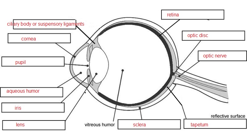
42 human eye with labels
All About the Eye Chart - American Academy of Ophthalmology You stand 20 feet away from the Snellen chart, and read from it without your glasses or contacts. You cover one eye and read out the smallest line of letters you can see. Then you cover the other eye and do it again. In some offices, you view the chart through a mirror. This means the test can be done with less than 20 feet of space. Fashion Label BOTTER: From the Caribbean to Paris | Miami Herald Under the unconcerned eye of the mass tourism, the fishing industry and the environmental issues. Giving life again to the coral reef is giving back to nature, giving back to the world." The ...
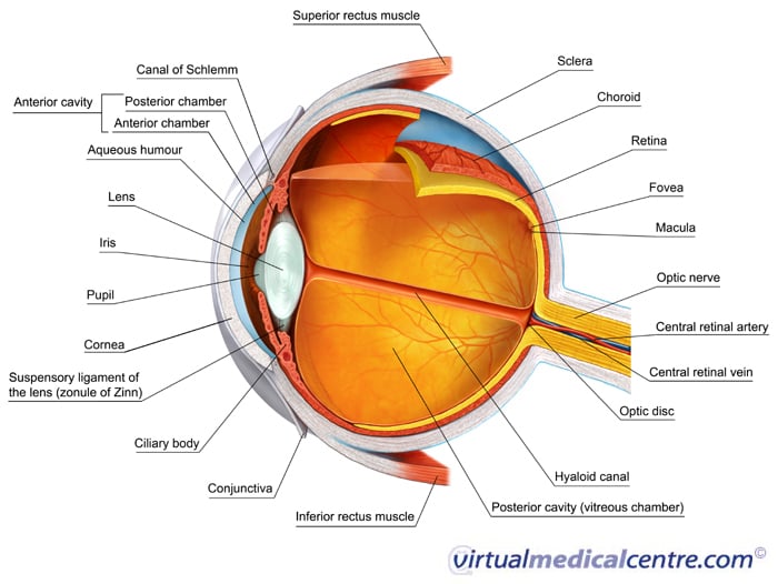
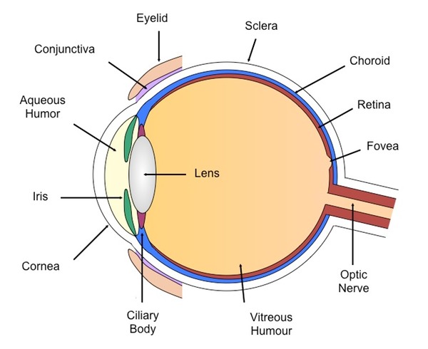
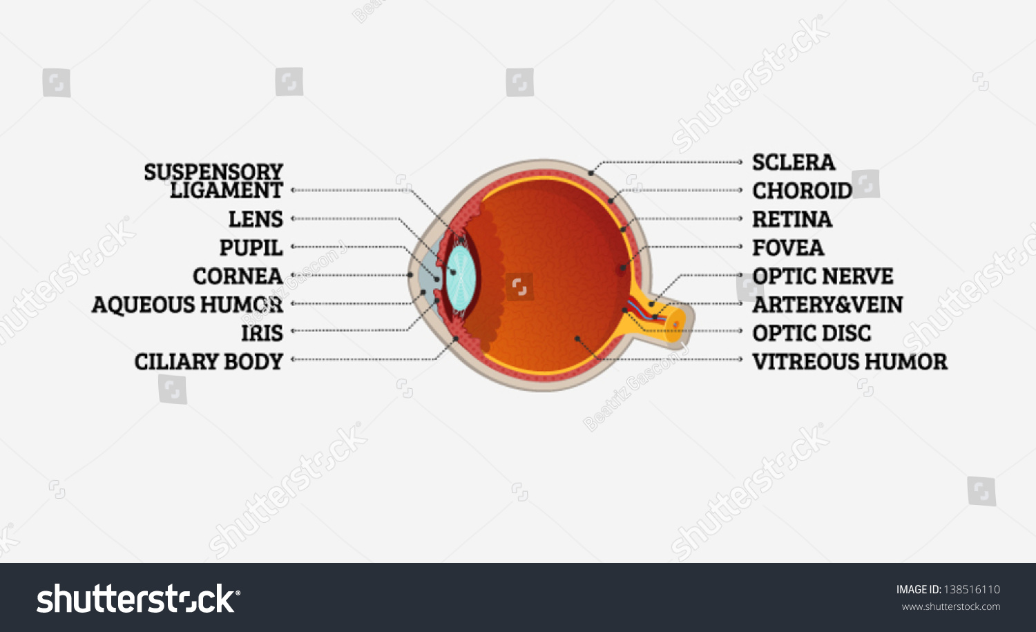
Eye Anatomy: 16 Parts of the Eye & Their Functions - Vision Center The following are parts of the human eyes and their functions: 1. Conjunctiva The conjunctiva is the membrane covering the sclera (white portion of your eye). The conjunctiva also covers the interior of your eyelids. Conjunctivitis, often known as pink eye, occurs when this thin membrane becomes inflamed or swollen.
Human eye with labels
Anatomy of the Eye - Verywell Health A cataract is a clouding of the lens and is a common occurrence that comes along with aging. Fortunately, cataracts grow slowly and may not affect your vision for several years. By age 65, over 90% of people have a cataract. Cataract treatment involves removing the cloudy lens surgically and replacing it with an implantable intraocular lens. Eye Anatomy and Physiology a Complete Detail - Study Read There are three major parts in each eye like The sclera (fibrous layer) Choroid layer Retina Eyes diagram showing the entire structure The sclera It makes up the outermost part of eye anatomy. It is made of a dense, strong fibrous wall consisting of the sclera that is 5/6 th and the cornea that is anterior 1/6 th of the eyeball. Invisible machine-readable labels that identify and track objects Invisible machine-readable labels that identify and track objects ... new smartphone model with a camera that utilizes the infrared (IR) range of the electromagnetic spectrum that the naked eye can't perceive. ... Objects Using Low-Cost Infrared-Based 3D Printing and Imaging Tools," is being presented at the ACM CHI Conference on Human ...
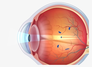
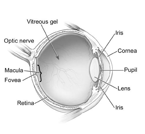
Human eye with labels. Anatomy of the Eye | BrightFocus Foundation Cornea: The outer, transparent structure at the front of the eye that covers the iris, pupil and anterior chamber; it is the eye's primary light-focusing structure. Drusen: Deposits of yellowish extra cellular waste products that accumulate within and beneath the retinal pigmented epithelium (RPE) layer. Fovea: The pit or depression at the ... Anatomy of the eye: Quizzes and diagrams | Kenhub Try our crash course in eye anatomy. One of our favorite ways to get to grips with all of the parts of the eye is by utilizing labeled diagrams. On a diagram of the eye, we can see all of the relevant structures together on one image. This helps us to understand how each one is situated and related to the other. Labeled diagram of the eye en.wikipedia.org › wiki › Human_penisHuman penis - Wikipedia The human penis is an external male intromittent organ that additionally serves as the urinary duct. The main parts are the root (radix); the body (corpus); and the epithelium of the penis including the shaft skin and the foreskin (prepuce) covering the glans penis . The Iris: Anatomy, Function, and Treatment - Verywell Health The iris sits in the uveal tract, which is the eye's middle layer. The iris lies in front of the lens and behind the cornea. It is made up of the following parts: 13. Iris pigment epithelium contains melanin granules and chromatophores that make up the eye color. Dilator and sphincter muscles that expand and contract to control the amount of ...
Understanding Different Eye Shapes: Which Do You Have? A shortened eyeball is known as hyperopia or farsightedness. This condition makes it difficult to see objects that are close. For example, it may be difficult to read a computer screen or a menu. When this condition is significant, the blurriness may also affect distance vision. In this case, the eyeball is shorter than what is considered normal. Parts Of The Eye Labeled Diagram Model And Their - SUNGLASSKY Parts of the eye-labeled diagram model are divided into three groups: the external outer layer, the middle layer, and the inner back layer. The outer layer is responsible for protecting the eye from environmental toxins and debris. The middle layer includes cells that allow light to enter and travel through the back layer to the retina. human ear | Structure, Function, & Parts | Britannica The human ear, like that of other mammals, contains sense organs that serve two quite different functions: that of hearing and that of postural equilibrium and coordination of head and eye movements. Anatomically, the ear has three distinguishable parts: the outer, middle, and inner ear.The outer ear consists of the visible portion called the auricle, or pinna, which projects from the side of ... towardsdatascience.com › swin-vision-transformersSwin/Vision Transformers — Hacking the Human Eye Jan 17, 2022 · 22k — 1K: This model is trained on Imagenet-22K consisting of 14 mil images and 22K class labels. It is then Fine-tuned on Imagenet-1K for a small number of epochs. We have seen this type of approach earlier in ViT. 224: This stands for the input image size. It is 224 x 224 across 3 channels for this model
Labeled imaging anatomy cases | Radiology Reference Article ... This article lists a series of labeled imaging anatomy cases by body region and modality. Brain CT head: non-contrast axial CT head: non-contrast coronal CT head: non-contrast sagittal CT head: angiogram axial CT head: angiogram coronal CT... Use of superordinate labels yields more robust and human-like visual ... Human visual recognition is outstandingly robust. People can recognize thousands of object classes in the blink of an eye (50-200 ms) even when the objects vary in position, scale, viewpoint, and illumination. What aspects of human category learning facilitate the extraction of invariant visual feat … Human Eye Lesson for Kids: Facts & Anatomy - Study.com Yes, it can but only through the aperture. Next, there is the lens. The light reflecting off objects being photographed must pass through the aperture, then through the lens. The amount of light... tinybop.com › apps › the-human-bodyThe Human Body educational app for curious kids by Tinybop ... Interactive labels in over 50 languages, including Chinese, Russian, German, French, Spanish, Japanese — and more! Meet the artist Designer and tinker Kelli Anderson created more than 200 drawings for The Human Body .
Eye Evolution
What's My Eye Shape? (Learn How to Tell Here) - Vision Center Elongated eyeballs are an indication of nearsightedness or myopia. This means the person has difficulty seeing far away. Most people with nearsightedness can see up close. But far-off items cause blurriness and trying to focus can lead to eyestrain and headaches. Nearsightedness is also caused by the cornea having an abnormal shape.

Human Eye Diagram Stock Illustration - Download Image Now ...
› photos › human-throat-anatomyHuman Throat Anatomy Pictures, Images and Stock Photos A set of human body parts and organ icons that include editable strokes or outlines using the EPS vector file. The icons include a human nose, ear, mouth, tongue ...

Our Eyes - Characteristics of a Human Eye - Turito
en.wikipedia.org › wiki › Human-readable_mediumHuman-readable medium - Wikipedia In computing, human-readable data is often encoded as ASCII or Unicode text, rather than as binary data. In most contexts, the alternative to a human-readable representation is a machine-readable format or medium of data primarily designed for reading by electronic, mechanical or optical devices, or computers.

Labelled Diagram Of Human Eye , Png Download - Label A Human ...
Microscope Types (with labeled diagrams) and Functions The working principle of a simple microscope is that when a lens is held close to the eye, a virtual, magnified and erect image of a specimen is formed at the least possible distance from which a human eye can discern objects clearly. Simple microscope labeled diagram Simple microscope functions It is used in industrial applications like:
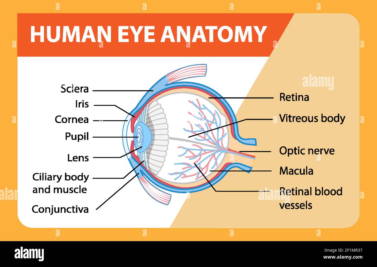
Diagram of human eye anatomy with label illustration Stock ...
Understanding Aqueous Humor and Vitreous Humor ... - NVISION Eye Centers The human eye is perhaps the most evolved and relied-upon part of the body. From the moment you wake up, you use your eyesight to accomplish most, if not all, of your daily tasks. ... Regular eye check-ups can help your doctor detect any issues with the aqueous humor and curate an adequate remedy. Various lifestyle choices can affect the ...

Solved: Label the diagram. Refer to Figure 43-18 to check ...
Diagram of Human Heart and Blood Circulation in It Four Chambers of the Heart and Blood Circulation. The shape of the human heart is like an upside-down pear, weighing between 7-15 ounces, and is little larger than the size of the fist. It is located between the lungs, in the middle of the chest, behind and slightly to the left of the breast bone. The heart, one of the most significant organs ...

External Anatomy of the Human Eye (With Labels)
Eye Diagram Quiz - ProProfs Quiz Try this amazing Eye Diagram Quiz quiz which has been attempted 5204 times by avid quiz takers. ... Label The Parts Of The Eye. People say that the eyes are the windows to a person's soul. ... How much did you get to understand about the human eye?... Questions: 8 | Attempts: 44923 | Last updated: Mar 22, 2022 . Sample Question. A is pointing ...

Human Eye- Parts of Human Eye | Turito
Quiz: Label The Parts Of The Eye - ProProfs Quiz Can you label the parts of the eye in the quiz below? Give it a try and evaluate yourself. The eye has many important parts, each with different functions, including the cornea, pupil, sclera, and many more. Can you tell where these parts are located and what function they perform?
FREE! - The Human Eye Labeling Activity (Teacher-Made)
› news › article-11226473Hollie Hughes labels Meghan Markle as horrible human Sep 19, 2022 · An Australian Liberal senator has unleashed a brutal attack on Meghan Markle and Prince Harry, saying she's sick of the pair and labelling them 'awful, revolting people.'. Hollie Hughes didn't ...
Anatomy - Vision
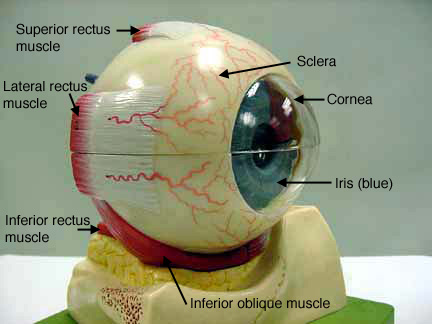
Anatomy and Structure of the Human Eye (With Diagrams) The sclera is the outermost layer of the eyeball. It is the white (and opaque) part. The muscles responsible for moving the eyeball are attached to it at the sclera. Cornea At the front of the eyeball, the sclera becomes the cornea—the transparent, dome-shaped part of the eyeball.
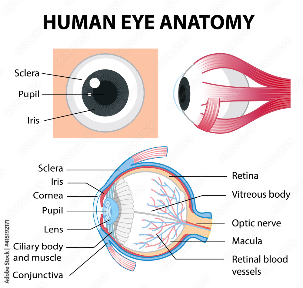
Diagram of human eye anatomy with label Stock Vector | Adobe ...
idlabelinc.com › common-types-warehouse-labelsCommon Types of Warehouse Labels - ID Label Inc. Warehouse Rack Beam Labels. The most common type of warehouse label is found on rack beams. These labels mark each rack bay location and are used to identify products for storing, picking and inventory management. They typically include a one- or two-dimensional barcode image and human-readable letters and numbers.
Free Blank Eye Diagram, Download Free Blank Eye Diagram png ...
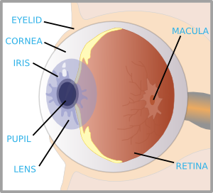
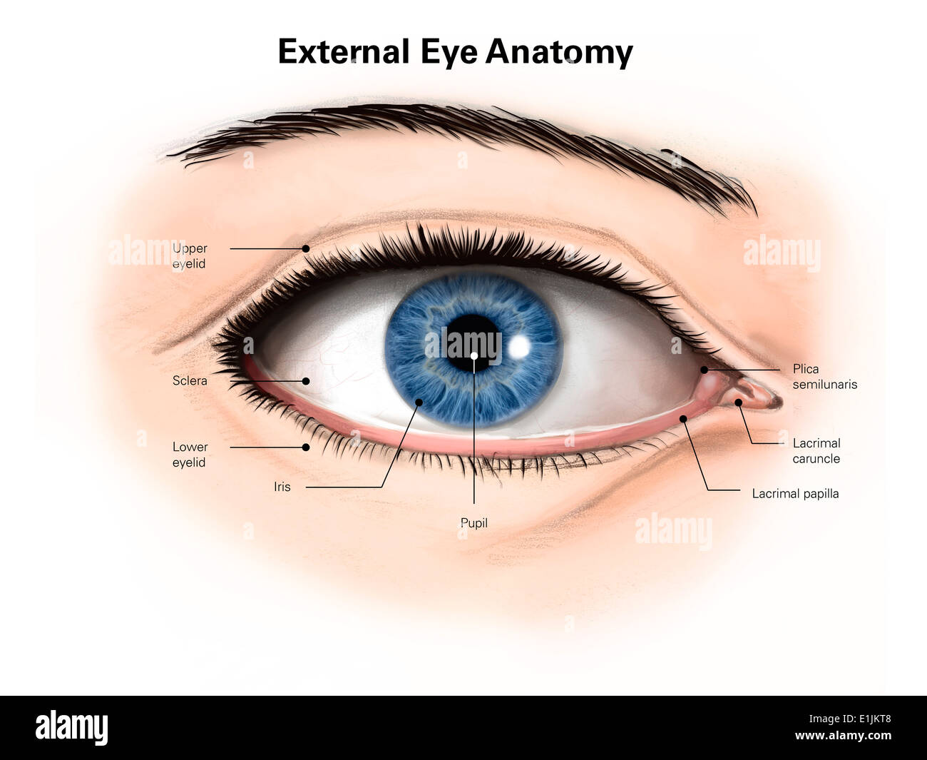
External and internal eye anatomy - MedlinePlus Overview. The cornea allows light to enter the eye. As light passes through the eye the iris changes shape by expanding and letting more light through or constricting and letting less light through to change pupil size. The lens then changes shape to allow the accurate focusing of light on the retina. Light excites photoreceptors that ...
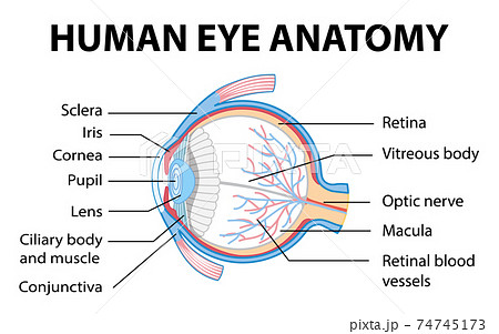
Diagram of human eye anatomy with label - Stock Illustration ...
What Eye Problems Look Like - WebMD It's called presbyopia, which means "old eye" in Greek. Most people start to notice it in their 40s. The eyes' lenses become less flexible and can't change shape to focus on objects at reading...

3d Image Render Of Diagram Of Eye Anatomy With Label For ...
Important Question for Class 10 Science Human Eye and Colourful World Excessive curvature of eye lens. Hypermetropia can be caused due to following reasons. Shortening of eyeball. Focal length of eye lens becomes too long. Presbyopia is caused due to gradual weakening of ciliary muscles and diminishing flexibility of eye lens due to ageing. Correction of these defects:

Draw a neat and label diagram of human eye and explain ...
Eyeball: Structure and function | Kenhub The eyeball consists of three layers; fibrous, vascular and nervous ( retina ). Functionally, the most important layer is the retina, which receives the external visual stimuli. The posterior pole of the eyeball is connected with the optic nerve (CN II), which conveys the information from the retina to the brain.
Eye With Labels Clip Art at Clker.com - vector clip art ...
Human Eye and Colourful World Class 10 Notes Science Chapter 11 Myopia (Short-sightedness): It is a kind of defect in the human eye due to which a person can see near objects clearly but he cannot see the distant objects clearly. Myopia is due to (i) excessive curvature of the cornea. (ii) elongation of eyeball.
Structure Of Human Eye Without Label Transparent PNG ...
Teeth Numbers and Names - Human Teeth Chart - Dayo Dental Teeth numbers 14 and 15 are your upper left molars. If you are getting cosmetic dentistry using veneers, you usually want to enhance the most visible part, teeth numbers 6 - 11 on the upper and 22 - 26 on the lower. For movie fans, vampires can extend their eye teeth (canines): 6, 11, 22 and 27. Teeth Numbers and Names

Schematic drawing of the human eye. Adapted from ...
Parts of Human Eye and Their Functions | MD-Health.com The different parts of the eye allow the body to take in light and perceive objects around us in the proper color, detail and depth. This allows people to make more informed decisions about their environment. If a portion of the eye becomes damaged, you may not be able to see effectively, or lose your vision all together. What are the parts ?

Label the Eye Flashcards | Quizlet
Invisible machine-readable labels that identify and track objects Invisible machine-readable labels that identify and track objects ... new smartphone model with a camera that utilizes the infrared (IR) range of the electromagnetic spectrum that the naked eye can't perceive. ... Objects Using Low-Cost Infrared-Based 3D Printing and Imaging Tools," is being presented at the ACM CHI Conference on Human ...

Cross-section of the human eye with labels - Stock ...
Eye Anatomy and Physiology a Complete Detail - Study Read There are three major parts in each eye like The sclera (fibrous layer) Choroid layer Retina Eyes diagram showing the entire structure The sclera It makes up the outermost part of eye anatomy. It is made of a dense, strong fibrous wall consisting of the sclera that is 5/6 th and the cornea that is anterior 1/6 th of the eyeball.

Label the Eye Quiz
Anatomy of the Eye - Verywell Health A cataract is a clouding of the lens and is a common occurrence that comes along with aging. Fortunately, cataracts grow slowly and may not affect your vision for several years. By age 65, over 90% of people have a cataract. Cataract treatment involves removing the cloudy lens surgically and replacing it with an implantable intraocular lens.

AP Human Eye Label Flashcards | Quizlet

External features of The Human Eye

Identify and label the parts of the human eye? Subject ...
External anatomy of the human eye (with labels Stock Photo ...
The Human Eye: A Diagram - FamilyConnect
0514 Anatomy Of Human Eye Medical Images For PowerPoint ...
Cow Eye Dissection
Human eye - Teaching resources
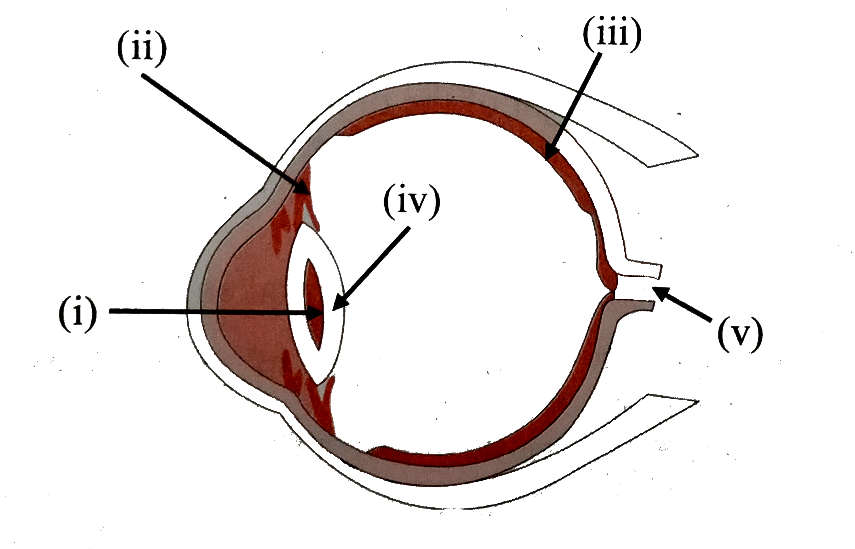
Label the parts of the following diagram of the human eye and ...

Labeled Eye Diagram | Science Trends
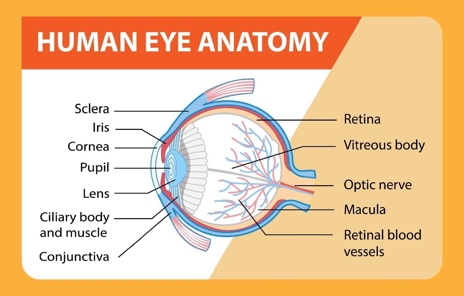
Diagram of human eye anatomy with label 1945551 Vector Art at ...

Quick Quiz
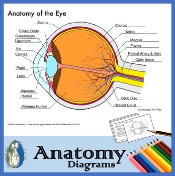
Anatomy of the Eye Diagrams for Coloring/Labeling, with ...
Eye Models
/GettyImages-695204442-b9320f82932c49bcac765167b95f4af6.jpg)
Structure and Function of the Human Eye
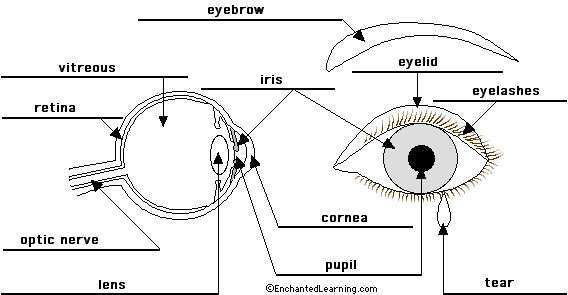
Eye Anatomy Diagram - EnchantedLearning.com

Module 1: Labeled Diagram of the Eye | Diagram of the eye ...
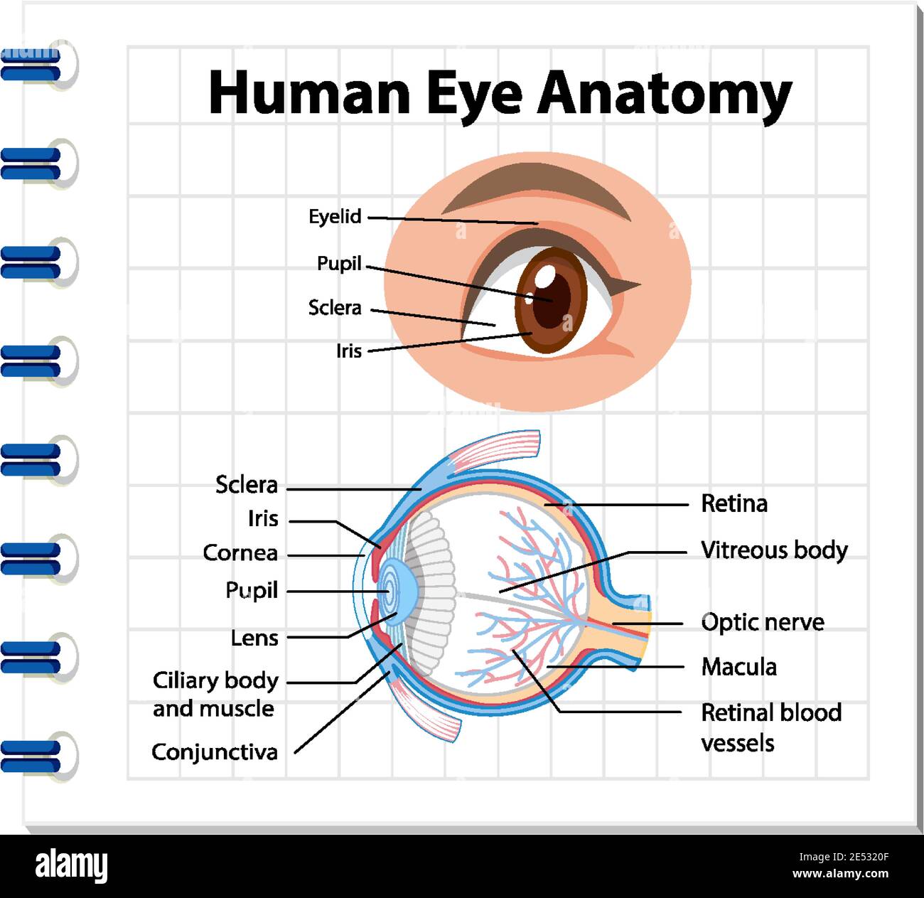
Diagram of human eye anatomy with label illustration Stock ...

Draw the struture of human eye and label its parts. - Science ...
Label Eye Printout - EnchantedLearning.com
Human Eye Crosssection Labeling Stock Vector (Royalty Free ...
Post a Comment for "42 human eye with labels"