38 heart structure with labels
Heart: Anatomy and Function - Cleveland Clinic Heart. Your heart is the main organ of your cardiovascular system, a network of blood vessels that pumps blood throughout your body. It also works with other body systems to control your heart rate and blood pressure. Your family history, personal health history and lifestyle all affect how well your heart works. Appointments 800.659.7822. Label Heart Anatomy Diagram Printout - EnchantedLearning.com | Heart ... Heart Coloring Pages Free Coloring Description This diagram shows the way blood flows through the heart. The areas of the heart with MORE oxygen are labeled with an "R". Students will color these areas RED. The areas of the heart with LESS oxygen are labeled with a "B". Students will color these areas BLUE.
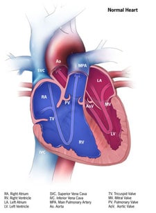
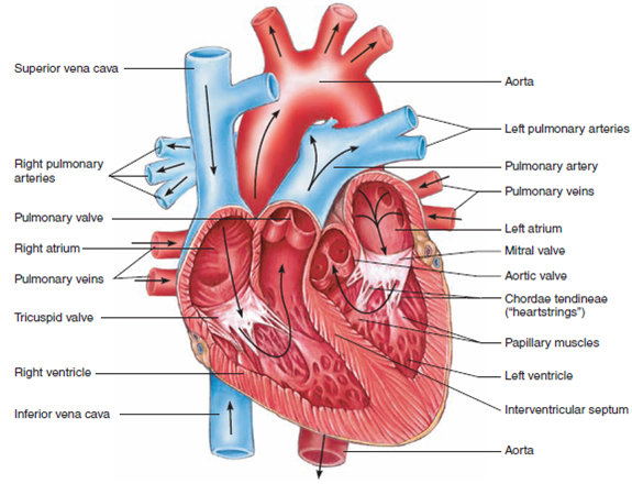
Heart Blood Flow | Simple Anatomy Diagram, Cardiac Circulation ... - EZmed Step 1 and 6 involve a blood vessel, which makes sense as this is how blood enters and exits that side of the heart. Steps 2-5 involve a chamber, valve, chamber, and valve. So if you remember this general pattern, it will help you recall the order in which blood flows through each side of the heart. Right Side of the Heart SVC/IVC Right Atrium
Heart structure with labels
The Anatomy of the Heart, Its Structures, and Functions The heart is the organ that helps supply blood and oxygen to all parts of the body. It is divided by a partition (or septum) into two halves. The halves are, in turn, divided into four chambers. The heart is situated within the chest cavity and surrounded by a fluid-filled sac called the pericardium. This amazing muscle produces electrical ... Heart Labeling Quiz: How Much You Know About Heart Labeling? Here is a Heart labeling quiz for you. The human heart is a vital organ for every human. The more healthy your heart is, the longer the chances you have of surviving, so you better take care of it. Take the following quiz to know how much you know about your heart. Questions and Answers 1. What is #1? 2. What is #2? 3. What is #3? 4. What is #4? Structure of the Heart | SEER Training Layers of the Heart Wall Three layers of tissue form the heart wall. The outer layer of the heart wall is the epicardium, the middle layer is the myocardium, and the inner layer is the endocardium. Chambers of the Heart The internal cavity of the heart is divided into four chambers: Right atrium Right ventricle Left atrium Left ventricle
Heart structure with labels. Label the heart — Science Learning Hub In this interactive, you can label parts of the human heart. Drag and drop the text labels onto the boxes next to the diagram. Selecting or hovering over a box will highlight each area in the diagram. Right ventricle Right atrium Left atrium Pulmonary artery Left ventricle Pulmonary vein Semilunar valve Vena cava Aorta Download Exercise Tweet 13+ Heart Diagram Templates - Sample, Example, Format Download This image can be used for text book representations of the interior labels of the human heart. Free Download. Color Heart Diagram Sample Format Free Download. ... thevirtualheart.org This structure of the heart along with the functions is available for download in the PDF format. This provides a clear explanation of the working of the human ... Labelling the heart — Science Learning Hub Labelling the heart — Science Learning Hub Labelling the heart Add to collection The heart is a muscular organ that pumps blood through the blood vessels of the circulatory system. Blood transports oxygen and nutrients to the body. It is also involved in the removal of metabolic wastes. Topics Concepts Citizen science Teacher PLD Glossary Sign in Label the HEART | Circulatory System Quiz - Quizizz True or False: Blood flows in the following sequence in the heart: Vena cava, right atrium, right ventricle, pulmonary artery, lungs, pulmonary veins, left atrium, left ventricle, aorta. Q. True or False: There are four chambers in the heart. Q. Place the pathway of blood through the heart in the correct sequence. Q.
How to Draw the Internal Structure of the Heart (with Pictures) Make sure to label the following: Superior Vena Cava Inferior Vena Cava Pulmonary Artery Pulmonary Veins Left Ventricle Right Ventricle Left Atrium Right Atrium Mitral Valves Aortic Valves Aorta Pulmonic Valve (Optional) Tricuspid Valve (Optional) 6 To finish, label "The Human Heart" above the sketch. Tips Use pencil The Anatomy of the Heart - Quiz 1 - Free Anatomy Quiz The circulatory system - lower body image, with blank labels attached. The circulatory system - a PDF file of the upper and lower body for printing out to use off-line. Describe and explain the function of the circulatory system - The circulatory system consists of the heart, the blood vessels (veins, arteries, and capillaries), and the blood. Label the Heart Diagram | Quizlet Start studying Label the Heart. Learn vocabulary, terms, and more with flashcards, games, and other study tools. Structure of Heart (With Diagram) | Circulatory System | Human Physiology The heart is consisting of three layers: 1. Pericardium or outer covering layer: The heart lies in a double membranous sac of pericardium with serous fluid between the two layers. This is known as pericardial fluid. By its lubricating action, the heart can move freely or contracts and expands without any injury.
Heart Diagram - 15+ Free Printable Word, Excel, EPS, PSD Template ... 99+ FREE & Premium Heart Drawings - Download NOW Beautifully Designed, Easily Editable Templates to Get your Work Done Faster & Smarter. Free Download Label The Parts Of The Heart depts.washington.edu | Having the heart diagram for studies or for scientific purpose has been made easy through this template. The structure of the heart - Structure and function of the heart ... The heart is a large muscular pump and is divided into two halves - the right-hand side and the left-hand side. The right-hand side of the heart is responsible for pumping deoxygenated blood to ... Diagram of Human Heart and Blood Circulation in It A heart diagram labeled will provide plenty of information about the structure of your heart, including the wall of your heart. The wall of the heart has three different layers, such as the Myocardium, the Epicardium, and the Endocardium. Here's more about these three layers. Epicardium Heart Diagram for Kids - Bodytomy As you can see in the diagram of the heart, that heart is divided in four chambers, namely, right atrium, left atrium, right ventricle and left ventricle. Each of the chambers is separated by a muscle wall known as Septum. The left side of the heart receives oxygen rich blood from the lungs and pumps it out the whole body.
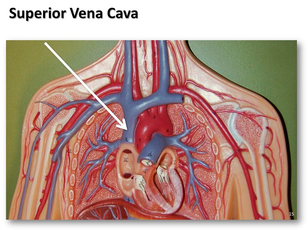
Superior vena cava - The Anatomy of the Veins Visual Guide… | Flickr
Structure of the Heart | The Franklin Institute The heart consists of four chambers: two atria on the top and two ventricles on the bottom. Looking at the Valentine's Day heart, the two rounded humps at the top are rounded like the top of a lower-case "a." The bottom is shaped like a "v." Feel it working What else is inside your heart?

Medical Encyclopedia - Structure: Structure of the Heart - Aviva | THE HEART | Pinterest | Heart ...
Heart anatomy: Structure, valves, coronary vessels | Kenhub Heart anatomy. The heart has five surfaces: base (posterior), diaphragmatic (inferior), sternocostal (anterior), and left and right pulmonary surfaces. It also has several margins: right, left, superior, and inferior: The right margin is the small section of the right atrium that extends between the superior and inferior vena cava .

Simple heart diagram to label by kpendlebury - Teaching Resources - TES
Simple heart diagram | Simple heart diagram labeled | Human heart ... Internal structure of human heart shows four chambers viz. two atria and two ventricles and couple of blood vessels opening into them. The wall of two ventricles are strong and sturdy when compared to atria. Before we start, we shall recall the basic proportions of heart and its chambers. The right Auricle is larger than left.
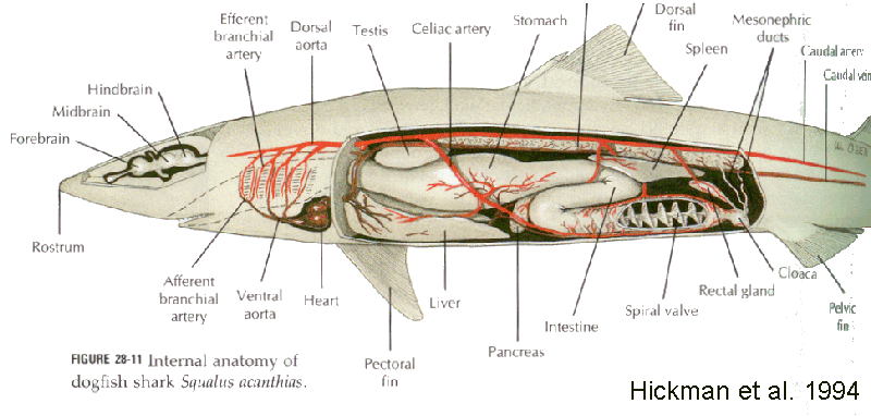
Chondrichthyes
Heart Anatomy: Labeled Diagram, Structures, Function, and Blood Flow There are 4 chambers, labeled 1-4 on the diagram below. To help simplify things, we can convert the heart into a square. We will then divide that square into 4 different boxes which will represent the 4 chambers of the heart. The boxes are numbered to correlate with the labeled chambers on the cartoon diagram.
How the Heart Works | Congenital Heart Defects | NCBDDD | CDC
Human Heart (Anatomy): Diagram, Function, Chambers, Location in ... - WebMD Human Heart (Anatomy): Diagram, Function, Chambers, Location in Body The right atrium receives blood from the veins and pumps it to the right ventricle. The right ventricle receives blood from the...

China U. S Trade War Heading To Economic Collapse : heading,News, breakingnews, globalnews ...
Structure and Function of the Heart - News-Medical.net The heart is a muscle whose working mechanism is made possible by the many parts that operate together. The organ is divided into several chambers that take in and distribute oxygen-poor or oxygen ...

Nucleus, the commanding centre of the cell ~ Biology Exams 4 U
Easy way to draw heart structure by 5 steps - YouTube My youtube channel : facebook page : way to draw hea...
😀 Drag the labels to identify structural components of the heart. Label the heart — Science ...
Heart Diagram with Labels and Detailed Explanation The heart is located under the ribcage, between the lungs and above the diaphragm. It weighs about 10.5 ounces and is cone shaped in structure. It consists of the following parts: Heart Detailed Diagram Heart - Chambers There are four chambers of the heart . The upper two chambers are the auricles and the lower two are called ventricles.

Loretta Hagen Sundown Till Dawn by Richard Cuccaro Original photo: Tracy Stoft Collage w/ sky ...
Human Heart Diagram Labeled | Science Trends List Of Heart Structures Heart Chambers Ventricles - The bottom two heart chambers. Atra - The upper two heart chambers. Wall Of The Heart Sinoatrial Node - A collection of tissue that releases electrical impulses and defines the rate of contraction for the heart. Atrioventricular Bundle - The fibers which transmit cardiac impulses.

Pin by Daffodilcooper on BSC2086 | Heart model, Anatomy models labeled, Cardiac anatomy
Heart Diagram with Labels and Detailed Explanation - BYJUS Diagram of Heart. The human heart is the most crucial organ of the human body. It pumps blood from the heart to different parts of the body and back to the heart. The most common heart attack symptoms or warning signs are chest pain, breathlessness, nausea, sweating etc. The diagram of heart is beneficial for Class 10 and 12 and is frequently ...

heart: Heart Labeled
Structure of the Heart | SEER Training Layers of the Heart Wall Three layers of tissue form the heart wall. The outer layer of the heart wall is the epicardium, the middle layer is the myocardium, and the inner layer is the endocardium. Chambers of the Heart The internal cavity of the heart is divided into four chambers: Right atrium Right ventricle Left atrium Left ventricle

Simplified Heart Labeled Decal | Shop Fathead Anatomical Images Graphics
Heart Labeling Quiz: How Much You Know About Heart Labeling? Here is a Heart labeling quiz for you. The human heart is a vital organ for every human. The more healthy your heart is, the longer the chances you have of surviving, so you better take care of it. Take the following quiz to know how much you know about your heart. Questions and Answers 1. What is #1? 2. What is #2? 3. What is #3? 4. What is #4?

Labeled Diagram Of The Heart Simple - Photos Idea
The Anatomy of the Heart, Its Structures, and Functions The heart is the organ that helps supply blood and oxygen to all parts of the body. It is divided by a partition (or septum) into two halves. The halves are, in turn, divided into four chambers. The heart is situated within the chest cavity and surrounded by a fluid-filled sac called the pericardium. This amazing muscle produces electrical ...
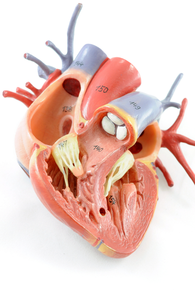
Label Heart Structure
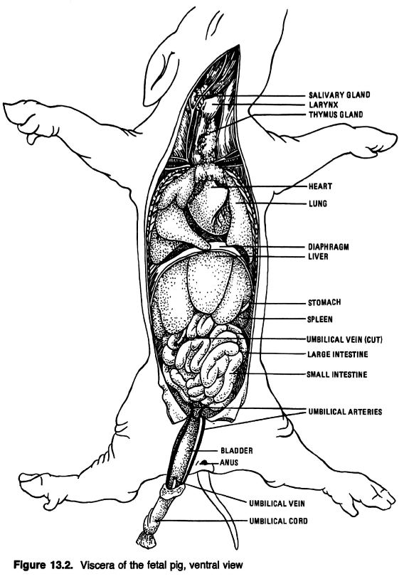
Anatomical Drawings of a Fetal Pig
32 Label The Diagram Of The Heart - Labels Database 2020
Post a Comment for "38 heart structure with labels"